Heart failure occurs when the heart cannot pump enough blood to meet the body’s demands. This decline in contractile function is caused by a variety of conditions but mainly following myocardial infarction, when blood flow to the heart is reduced.
ECG, Blood pressure and Blood flow investigation
The investigation of ECG, blood pressure and flow recordings and the study of heart contractility-relaxation allow researchers to detect and understand cardiac structural abnormalities which enhance the heart failure risk. A set of techniques including in vivo and ex vitro analysis can be considered (Fenske et al, 2016).
Interaction between cardio and pulmonary outcomes
When the left ventricle is weakened, pressure increases in the lung vasculature allowing fluid to collect (pulmonary edema). When the right ventricle is weakened, it is often a result of pulmonary hypertension, where the pulmonary arteries are narrowed or damaged. This makes it harder for blood to flow through the lungs and forces the right ventricle to work harder, eventually weakening the muscle.
Understanding this close interaction requires a two-step approach, investigating both cardio and pulmonary outcomes.
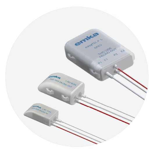
easyTEL+ system allows for simultaneous acquisition of ECG and pressure data in conscious rodent to large animals to understand the mechanism related to Heart Failure during chronic studies without confounding effects of surgery or anesthesia.
Different models can be developed such as Atrial Fibrillation. Heart failure (HF) and atrial fibrillation (AF) are two closely inter-related conditions with similar risk factors and share physiopathology that are likely to dominate the next 50 years of cardiovascular care.
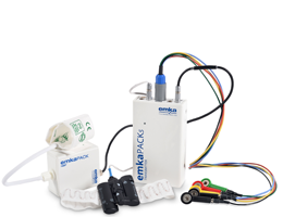
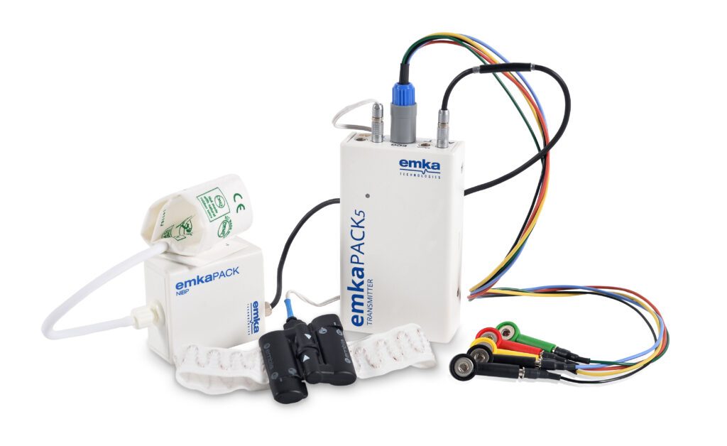
emkaPACK5 system allows for simultaneous acquisition of ECG and respiratory data in large animals to understand the relationship of cardiovascular and pulmonary outcomes without confounding effects of surgery or anesthesia.
When subjects are particularly fragile and cannot tolerate surgery or anesthesia, as with cardiomyopathy or pulmonary hypertension, emkaPACK5 is a good alternative to implanted telemetry.
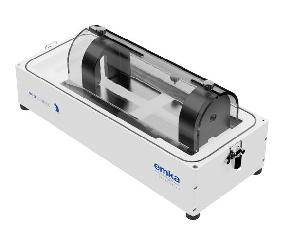
ecgTUNNEL benefits of an innovative design allowing for simultaneous acquisition of ECG and respiratory data in small animals, to understand the relationship of cardiovascular and pulmonary outcomes without confounding effects of surgery or anesthesia.
When subjects are particularly fragile and cannot tolerate surgery or anesthesia, as in cardiomyopathy disease, ecgTUNNEL is a good alternative to implanted telemetry.
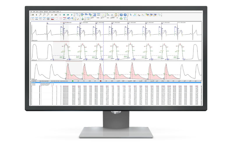
ecgAUTO allows for simultaneous analysis of ECG and blood pressure (arterial and left ventricular) from rodents to large animals. Add-on modules utilize user-defined parameters for advanced HRV and baroreflex sensitivity (BRS) analysis to study risk factors for sudden cardiac arrest and disease progression of heart failure.
A respiratory module allows ecgAUTO to serve as a single platform for interdisciplinary studies.
The investigation of ECG and blood pressure recordings and the study of heart contractility-relaxation allow researchers to detect and understand these cardiac structural abnormalities, which enhance the heart failure risk.
With the isolated heart setup, the researchers can acquire Left Ventricular Pressure, which is analyzed in IOX2 software to get many parameters such as systolic and diastolic pressure and others. Indeed, the systolic pressure is a clinically important determinant of LVP fall in cardiac overload and in congestive heart failure (Gillebert et al, 1997).
In addition, ECG can be captured through two electrodes placed at the surface of heart and analyzed in ecgAUTO software.
In the working heart perfusion mode, the flow of perfusate mimics the flow of blood in situ. As the name implies, this technique allows the heart to perform its physiological pumping action, i.e. it performs pressure/volume work. Therefore, it provides a complete analysis of heart function and can be used for the study of ischemia-reperfusion. The experiments can even be continued when a cardiac arrest occurs.
The Langendorff perfused heart experiments can be useful in documenting the structural and functional deterioration in animal models of heart failure and the effect of therapies on the transition from compensated to decompensated heart failure. Another possibility is the study of substrate preference using metabolomic (glucose, fatty acids) in heart failure models.
Cardiovascular remodelling including heart and blood vessels is observed during heart failure.
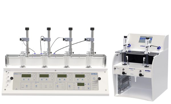
In order to understand the mechanisms of these regulatory processes in health and disease, investigations into the functioning of these vessel segments, i.e. of their reactions to the application of external stimuli such as vasoactive substances, pressure, etc. is performed in tissue baths or myographs.
The goal of these studies is to look at the response of the vessel to agonists or antagonists added to the bath, or to study the inotropic or lusitropic responses, generating a Concentration Response Curve (CRC).
Rat aortic rings or mesenteric artery are widely used for the study of hypertension or atherosclerosis, just to cite a couple.
The impact of diabetes and obesity on organ dysfunction and in the occurrence of cardiovascular diseases is also widely studied with tissue baths, as well as dromotropic response on right atria or papillary muscle.
The development of genetic manipulation has extended the scope of isolated organ baths. Transgenic mice overexpressing genes can be subjected to organ baths experiment in order to characterize the pharmacological effect of this manipulation.
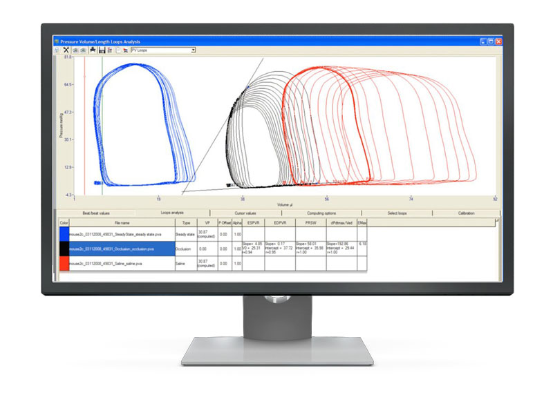
Real-time left ventricular (LV) or Right ventricular (RV) pressure–volume loops provide a framework for understanding cardiac mechanics in experimental animals and humans. Such loops can be generated by real-time measurement of pressure and volume within the left or right ventricle. Several physiologically relevant hemodynamic parameters such as stroke volume, cardiac output, ejection fraction, myocardial contractility, etc. can be determined from these loops.