Ischaemic heart diseases continue to be a major global health concern and a leading cause of death. To develop more effective treatments, it is essential for researchers to work with animal models that closely replicate human cardiac physiology. In a notable step forward, Pius et al. present a refined, minimally invasive ovine model of ischaemia–reperfusion–infarction which incorporates implantable defibrillators and addresses many limitations seen in previous approaches, enhancing the safety, reproducibility, and clinical relevance of preclinical cardiac research.
Traditional large-animal models for myocardial infarction (MI) often involve highly invasive procedures and carry a significant risk of mortality, limiting both the ethical feasibility and translational value of the research. In their study, Pius et al. address these challenges by developing a more refined approach that prioritizes both animal safety and model reproducibility. At the core of the methodology is a catheter-based balloon angioplasty, used to temporarily occlude the left anterior descending (LAD) artery before reperfusion. This controlled ischaemia–reperfusion injury effectively simulates the pathophysiological conditions of a human MI, but without the need for open-chest surgery. A key innovation in this model stands in the use of implantable cardioverter defibrillators (ICDs), which were placed prior to MI induction. This enables real-time monitoring and correction of potentially fatal ventricular arrhythmias, dramatically lowering the high intra-operative mortality associated with previous models. In addition, a carefully designed prophylactic anti-arrhythmic regimen—comprising amiodarone, lidocaine, and atenolol—was used to further mitigate the likelihood of arrhythmic events during the procedure.
The refined model achieves a notable reduction in intra-operative mortality to 6.7%, a significant improvement compared to previous models reporting rates between 13% and 43%. Validation of the model shows that it effectively reproduces the key features outlined in the fourth universal definition of myocardial infarction.
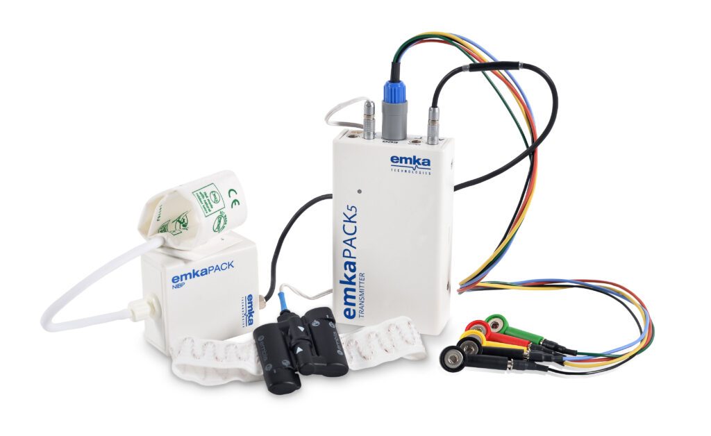
Throughout the study, electrocardiograms were recorded non-invasively using the emkaPACK telemetry system equipped with 5 electrodes. The ECG signal was acquired through IOX software, displaying characteristic ST-segment elevations and T-wave changes. Concurrently, cardiac biomarkers, notably cTnI, showed a typical rise and fall pattern, confirming myocardial injury. Echocardiographic assessments further supported these findings, showing regional wall motion abnormalities consistent with infarcted myocardial tissue. These indicators collectively affirmed that the ovine model accurately mirrored the clinical and physiological hallmarks of MI.
The work by Pius et al. presents a major advancement in translational cardiac research. By refining the ischaemia–reperfusion–infarction model in sheep and enhancing it with safety mechanisms like implantable defibrillators, they have established a robust, reproducible, and clinically relevant tool for studying myocardial infarction and testing future therapies.