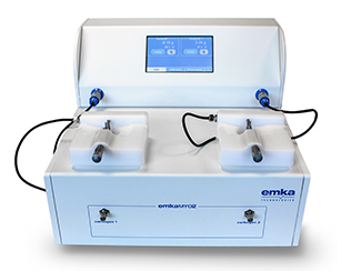
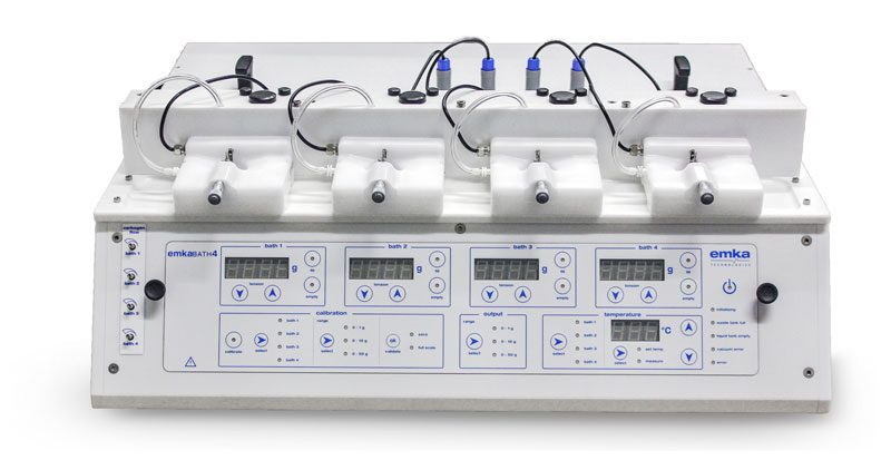
Our myograph systems are used to investigate the functional responses and vascular reactivity of small arteries, veins, lymph vessels and tracheal smooth muscles, with lumen diameter from 100µm to 3mm and segment lengths up to 3.5mm.
Wire myography is an ex vivo technique widely used in preclinical research, to examine microvessels contractility following drug challenges. It is commonly used in drug development and toxicology studies, to look at the response of the vessel from a wide range of species to agonists or antagonists added to the bath, generating a Concentration Response Curve (CRC).
| Specifications | emkaMYO2 | emkaMYO4 |
|---|---|---|
| Max. number of baths | 2 | 4 |
| Bath volumes | 5 ml | 5 ml |
| Optional tubular tissue baths | ||
| Microvessel size | Diameter: 100μm-3mm / Length: 3.5 mm max | |
| Bath heating regulation | Individual temperature by bath | Same temperature for all baths |
| Physiologic liquid heating |
No in line heating.
Water bath required. |
In line heating of the liquid before entering the bath |
| Bath temperature range | 20.0-39.0°C ± 0.1°C (68-102.2°F ± 0.18°F) | |
| Automatic filling | ||
| Automatic emptying | ||
| Frame size | 300x335x206 mm | 641x500x345 mm |
| Weight | 2 kg | 18 kg |
| Renewed solution by overflow | ||
| Automatic cleaning | ||
| Integrated amplifier | ||
| Bubbling | Frit glass | Frit glass |
| Analog output | 0-5 V | 0-5 V |
| Manual tissue tension | ||
| Connection | USB or analog | USB or analog |
| Protocols | ||
| Normalization | ||